Acoustic Neuroma Explained: Causes, Symptoms, And Treatments
An acoustic neuroma is a rare type of brain tumor (per the Acoustic Neuroma Association). Acoustic neuromas got their name because doctors and scientists originally thought that the tumors grew from the acoustic nerve, though later research showed that they come from Schwann cells of the vestibular nerve. Therefore, vestibular schwannoma is a more appropriate name, but the term acoustic neuroma is more common.
Schwann cells form a sheath around nerves, insulating them and increasing nerve impulse conduction (via the National Library of Medicine). Acoustic neuromas form when Schwann cells surrounding the vestibular nerve form a benign, or non-cancerous tumor, according to the Acoustic Neuroma Association. The vestibular nerve is "responsible for both hearing and balance" (per Healthline), so the first signs of an acoustic neuroma are typically problems with hearing function and balance.
These tumors tend to be slow growing, although they grow rapidly in some cases. Acoustic neuromas do not spread throughout the body but because they press on the brain as they grow, they can cause symptoms and health problems. Let's take a closer look at the causes, symptoms and available treatments.
Causes of acoustic neuromas
Acoustic neuromas are rare, affecting between 1 and 3.5 out of 100,000 people each year. According to a 2020 review in the journal Head and Neck Pathology, acoustic neuromas account for 8% of all intracranial tumors, or tumors that grow inside the skull, each year.
According to the Acoustic Neuroma Association, 95% of acoustic neuromas are spontaneous. These spontaneous tumors may arise from a random mutation on chromosome 22, specifically in a gene known as NF2 (per the review in Head and Neck Pathology). This gene is a tumor suppressor gene, meaning that it prevents tumors from forming. Mutations here deactivate the tumor suppressor gene, allowing tumors to form. Mutations in the tumor suppressor genes LZTR1, SMARCB1, and COQ6 "are also linked to schwannoma development." These mutations spontaneously arise in the cells of the patient and are not passed on from generation to generation.
Spontaneous cases of acoustic neuroma tend to be unilateral, affecting just one ear. In contrast, bilateral acoustic neuromas, or tumors that affect both ears, are more common in people with a condition called neurofibromatosis type 2 (NF2). NF2 explains the other 5% of acoustic neuroma cases that are not spontaneous.
Neurofibromatosis Type 2 can cause acoustic neuromas and other tumors
A 2021 review in the International Journal of Molecular Sciences describes neurofibromatosis type 2 (NF2) as a genetic disorder caused by a mutation in the NF2 gene, which encodes a tumor-suppressing protein known as Merlin. The mutation causes the NF2 gene to lose its tumor-suppressing function, and so the NF2 disorder is characterized by the formation of tumors in the "central and peripheral nervous system." Roughly one in 25,000 people have NF2.
People with NF2 are more likely to develop acoustic neuromas and they develop these tumors at an early age. By age 20, roughly 90% of patients with NF2 develop "bilateral vestibular schwannomas." These tumors may recur even after treatment. Half of NF2 patients will also develop meningiomas, which are tumors that form from the meninges — "the membranes that surround the brain and spinal cord" (per the Cleveland Clinic). Many patients also develop spinal schwannomas, a type of spinal cord tumor, according to the Mayo Clinic, ependymomas, a type of tumor in the brain or spinal cord (via the National Cancer Institute), and dermal schwannomas, a type of tumor of the nerve sheath that forms beneath the skin, explains a 2010 case report in the American Journal of Dermatopathology.
Because people with NF2 develop so many types of tumors at an early age, they require a team of specialists to manage their condition. This team may include family physicians, neurogeneticists, neuro-oncologists, neuro-otologists, neurosurgeons, neuro-ophthalmologists, neuroradiologists, social workers, and audiologists — along with speech, physical, and occupational therapists.
Risk factors for developing an acoustic neuroma
While NF2 is a serious risk factor for developing an acoustic neuroma, it is not the only one. In fact, NF2 only explains 5% of acoustic neuroma cases. The other 95% have a different root cause, including some environmental factors. A 2016 review published in the Yonsei Medical Journal identified leisure noise as a risk factor for acoustic neuromas. Exposure to leisure noise, such as listening to loud music or listening to music through earbuds or headphones, was associated with acoustic neuromas, perhaps by mechanically damaging the vestibular nerve and the surrounding tissue.
Another risk factor is exposure to radiation. Children who were exposed to radiation, typically as a treatment to reduce their tonsils or adenoids, were more likely to develop acoustic neuromas (per a 2008 study in the journal Neuro-Oncology). Survivors of the atomic bombs dropped on Hiroshima and Nagasaki were also more likely to develop these tumors, according to a 2002 study published in the Journal of the National Cancer Institute. These two studies demonstrate the radiation can lead to acoustic neuromas.
Interestingly, smoking reduces the risk of developing acoustic neuromas (per the 2016 review in the Yonsei Medical Journal). Researchers speculate that nicotine may have a protective effect against acoustic neuroma tumors or that smoking may starve the tumor cells of oxygen. However, the health risks of smoking outweigh any protective benefits.
Symptoms of an acoustic neuroma
Acoustic neuromas are slow-growing tumors, so their symptoms may take years to appear, according to the Mayo Clinic. When they do show up, these symptoms are easy to miss. As the tumor grows, it will start to press on nearby nerves, blood vessels, and muscles, making symptoms more apparent.
Because the vestibular nerve affects hearing, many symptoms of an acoustic neuroma involve hearing. Hearing loss on one side is the most common symptom of an acoustic neuroma. Patients may also experience a feeling of fullness in the affected ear. Others develop tinnitus, which is a ringing, roaring, clicking, buzzing, or hissing sound in one or both ears, according to the National Institute on Deafness and Other Communication Disorders.
The vestibular nerve also plays a role in balance, so patients with acoustic neuromas may feel unsteady or lose their sense of balance and develop vertigo. Pressure from the tumor on other nerves or muscles may lead to facial numbness or weakness, loss of muscle movement, difficulty swallowing, headaches, a feeling of pressure in the head, a change in taste, and a change in tear production.
According to Johns Hopkins Medicine, death is a rare but potential outcome of an acoustic neuroma. Untreated tumors can "compress the cerebellum and brain stem" or cause an increase of fluid on the brain.
How to diagnose an acoustic neuroma
The symptoms of an acoustic neuroma can look like the symptoms of other middle and inner ear conditions (per Johns Hopkins Medicine), so it is critical that doctors recommend the right testing for an appropriate diagnosis. In addition, early diagnosis of an acoustic neuroma can improve long-term health outcomes.
Your doctor will typically start by asking you questions about your symptoms, then examining your ear. The doctor will then recommend further tests such as a hearing exam and imaging tests (per the Mayo Clinic).
According to Johns Hopkins Medicine, an audiologist will conduct a variety of hearing tests that assess how well you can hear sounds and how well you can distinguish between different sounds. These tests may assess one ear at a time. Your testing may look at an increased pure tone average to measure how loud a sound must be before you hear it, an increased speech reception threshold to measure how loud speech must be before you hear it, and decreased speech discrimination to measure how many words you can detect.
Your doctor will most likely recommend an MRI if your hearing tests indicate an acoustic neuroma. This imaging scan allows your doctor to visually confirm the presence of a tumor.
Other conditions mimic the symptoms of an acoustic neuroma
Proper testing and diagnosis are necessary to rule out other conditions with symptoms similar to symptoms of an acoustic neuroma. The National Organization for Rare Disorders provides information about conditions, such as Bell's palsy, Meniere's disease, and meningioma, that can look like an acoustic neuroma.
Bell's palsy is a neurological disorder that affects the seventh cranial nerve. It is non-progressive, meaning that it does not get worse with time. There is no known cause of Bell's palsy, although it may be linked to a viral infection, an immune disorder, or family history. Bell's palsy causes a sudden onset of facial paralysis, which may look like the facial weakness sometimes caused by an acoustic neuroma.
Meniere's disease affects the inner ear and can cause episodes of vertigo or dizziness, low-frequency hearing loss, tinnitus, and a feeling of fullness or pressure in the ear, all symptoms of an acoustic neuroma. People with Meniere's disease may experience symptoms daily or occasionally, although hearing loss and tinnitus can become permanent.
Meningiomas are benign tumors of the meninges, which is the membrane lining the brain and spinal cord. Meningiomas can occur anywhere in the brain or spinal cord, but if they develop near the nerves affecting balance and hearing they can cause the same symptoms as an acoustic neuroma. Common symptoms include hearing loss, dizziness, problems with balance, facial numbness, and difficulty swallowing.
There are several treatment options for acoustic neuroma
According to a 2020 review published in the journal Neuro-Oncology, patients have several options when it comes to treating an acoustic neuroma. Doctors may recommend different treatment strategies based on the tumor's size, morphology or shape, and the patient's comorbidities and preferences. Treatment options include observational management, stereotactic radiosurgery, or other surgical options.
Observational management is also known as watch and wait. Here, the doctors will monitor the tumor for growth and monitor the patient for hearing loss and speech discrimination. It is a good choice for patients who will comply with follow-up testing. When compared to patients treated with surgery or stereotactic radiosurgery, patients with conservative management had better facial and hearing nerve outcomes. They also reported a better quality of life when compared to patients who underwent surgery.
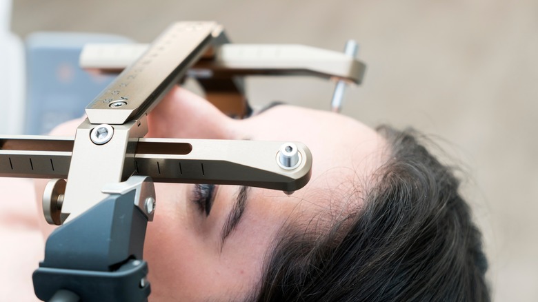
Stereotactic radiosurgery uses a large dose of irradiation to selectively treat the acoustic neuroma without damaging nearby tissue. Research suggests that it works best on acoustic neuromas that are small or medium in size. For tumors under 3 centimeters in size, it is the preferred treatment in terms of preserving facial nerve and hearing function.
There are three main surgical options: the middle fossa approach, the retrosigmoid approach, and the translabyrinthine approach. Surgeons will advise the patient on these options based on the patient's current hearing status, tumor characteristics, patient's preference, and the surgeon's expertise. The surgeon's experience has a large effect on surgery's outcome, so patients should consider choosing a high-volume center where acoustic neuroma surgery is performed regularly.
Surgical options to treat an acoustic neuroma
Your doctor will advise you on the best surgical approach based on the size of your tumor and its location. Here is more information from the Cleveland Clinic about surgical options.
With the middle fossa approach, the surgeon removes the tumor from the inner ear through an incision in the side of your head. This approach works well for small tumors inside the bony ear canal. It may preserve hearing and facial nerve function.
With the retrosigmoid approach, the surgeon makes an incision behind the ear to remove the tumor from the inner ear canal and a part of the skull called the cranial fossa. This approach works best for medium to large tumors that are pressing on the brain. It may preserve hearing and prevent facial paralysis.
With the translabyrinthine approach, "The surgeon makes an incision behind the ear, removing bones of the inner ear for a better view of the tumor" (via the Cleveland Clinic). This process causes total hearing loss on the affected side, so surgeons typically use this approach on patients who have already lost their hearing. This approach can be used for tumors of any size. While it does cause hearing loss, it preserves facial nerve function.
Your surgical team will attempt to remove the entire tumor but may choose to leave some of the tumor behind to protect the facial nerve. According to a 2020 review published in the journal Neuro-Oncology, residual tumors can lead to recurrence of the acoustic neuroma.
Complications from acoustic neuroma surgery
Any surgery comes with the risk of complications, and acoustic neuroma surgery is no different. Damage to your facial nerve function and hearing are two main complications from this surgery. No matter which surgical approach your team uses, they will monitor your facial nerve function and hearing throughout the surgery (per the Cleveland Clinic) to increase the chances of preserving them. Tumor size affects the chances of hearing loss. People with small to medium tumors who still have good hearing before the surgery have a 50% chance of retaining their hearing post-surgery.
There are other complications related to surgery to remove an acoustic neuroma. Complications related to the ear include tinnitus, dizziness, and problems with balance. Other complications such as dry eye, double vision, and difficulty closing the eyelid, affect the eye. Some people experience complications with the mouth such as dry mouth, problems with taste, and difficulty swallowing. Other surgical complications include cerebrospinal fluid leaks, infection, and headaches. Lastly, the tumor may grow back after the surgery.
Special considerations for patients with NF2
Patients with NF2 often develop acoustic neuromas on both sides, which can lead to complete deafness. Because of this risk, observational management is not the best approach for patients with NF2. The Acoustic Neuroma Association recommends that these patients work closely with their team of specialists to preserve their hearing for as long as possible.
The current recommended strategy for NF2 patients is to remove the smaller of the two tumors first, because removing smaller tumors typically preserves hearing on that side. If the surgery works and preserves hearing, patients should have the other tumor removed. If the surgery does not preserve hearing, observational management should be used on the other tumor to avoid losing hearing in both ears. However, if the second tumor continues to grow or starts to damage hearing on that side, it should be removed as well.
While stereotactic radiosurgery is an option, it is not always the best choice for NF2 patients. The cell type of acoustic neuromas in NF2 patients can be resistant to radiation and more likely to become malignant.
Emerging treatments for acoustic neuromas
Researchers continue to develop new treatment approaches for acoustic neuroma, particularly approaches that may allow patients to avoid surgery. A 2020 review in the journal Head and Neck Pathology discusses targeted biologic therapies that provide a pharmaceutical approach to treating acoustic neuromas.
One such therapy is the drug Bevacizumab, a monoclonal antibody. Monoclonal antibodies are proteins that look like immune system proteins that the body makes and can destroy specific targets like tumors in the body (per the Cleveland Clinic). Studies show that Bevacizumab can reduce tumor size, prevent growth, and improve hearing.
Another promising drug is Lapatinib, which inhibits EGFR and ErbB2, two growth factors involved in tumor growth (per a 2002 study in the journal Laboratory Investigation). A clinical trial showed that it helped reduce tumor volume and improved hearing in some participants.
Aspirin may also delay growth of acoustic neuromas. Studies have indicated that people with acoustic neuromas taking aspirin for other reasons had reduced tumor volume.
What to expect after treatment for an acoustic neuroma
According to the Cleveland Clinic, most patients can expect to stay in the hospital for two to three days after undergoing surgery to remove an acoustic neuroma. In the days and weeks following the surgery, patients will need to go through several tests. Your doctor may recommend an MRI or other imaging scans to confirm tumor removal and monitor the area. They will also order tests to see how the surgery affects your hearing, facial nerve function, and balance.
A 2022 study in the Journal of Clinical Neuroscience found that frail patients, defined as patients with exhaustion, low physical activity, weakness, slowness, and shrinking, experience worse outcomes after surgery. They are more likely to experience complications such as post-operative infection, facial paralysis, urinary tract infections, and hydrocephalus. They also tend to have longer hospital stays and higher rates of readmission.
Patients who have trouble with balance may need to go through vestibular rehabilitation therapy (VRT). According to the Vestibular Disorders Association, VRT is an exercise-based therapy performed by a specialized occupational or physical therapist. It can help patients through habituation, gaze stabilization, and balance therapy. Habituation reduces dizziness caused by quick movements, changes in posture, or visual stimuli. Gaze stabilization helps patients by improving their eye control. Balance training provides patients with customized exercises to help reduce their risk of falling and improve their ability to walk on uneven surfaces.
Restoration of hearing loss after an acoustic neuroma
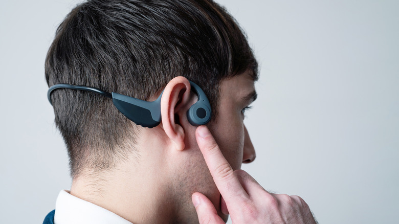
Patients who lose their hearing because of their acoustic neuroma and its removal often want to restore their hearing. Currently, there are a few options that may restore hearing: contralateral routing of sound aid, bone conduction hearing implant, and auditory brainstem implant (per the Cleveland Clinic). Talk to your doctor and audiologist about the best option for you.
A contralateral routing of sound (CROS) aid uses a microphone-like device to send the sound from the affected ear to the functional ear. This type of hearing aid is an external device.
A bone conduction hearing implant is surgically implanted in the skull behind the affected ear. The implant sends the sound from the affected ear to the functional ear.
The auditory brainstem implant also requires surgery. Here, the surgeon implants the device directly on the brain on its hearing centers. An auditory brainstem implant is a good option for patients with NF2.


